A clinical case that explains the technique step by step
Root perforation repair has historically been a treatment with a low success rate; however, recent techniques and materials utilized in root perforation repair, have dramatically improved the prognosis of both surgical and non surgical procedures. Root perforation is defined as an artificial communication between the root canal system to the supporting tissues of teeth often caused by using rotary burs inside root canals.
In my practice, I have found lot of perforations caused by an inappropriate post space preparation for permanent restoration of endodontically treated teeth. They are located in the middle part of the canal and, according to my personal statistic, 80 per cent of the cases involve the first lower molar: considering this tooth, 60 per cent of perforations are in the mesial root and 40 per cent in the distal root and they are always generated by an over preparation of post space that has not taken into consideration the geometry of the cross-sectional anatomy of the lower first molar. Another consideration is that large sized perforations may not respond to repair as well as smaller ones.
Diagnosis
Bacterial infection emanating either from the root canal or the periodontal tissues, or both, prevents healing and brings about inflammatory sequels where exposure of the supporting tissues is inflicted. Thus, painful conditions, suppurations resulting in tender teeth, abscesses, and fistulae including bone resorptive processes may follow (1). A narrow isolated periodontal defect is a possible sign of root perforation. To determine locally isolated vertical bone losses, periodontal probing should be carried out by walking the probe around the tooth while pressing gently on the floor of the sulcus (2).
Fig 1: In the first lower molar an X-ray can often show a bone loss between roots and diagnosis is easier than other teeth.
Fig 1: The MTA sandwich technique is the author’s personally recommended repair method for this kind of perforation and it has been well described by Fabio Gorni in this article.
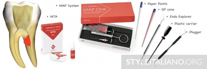
But what’s the equipment?
1) MTA is my choice material for perforation repair and numerous case reports exist in the literature showing excellent healing results with MTA.
2) The MAP (Micro-Apical Placement) System, provides an efficient method for placing repair materials and is the only one device that lasts over time. It has lot of tips, steel and NiTi, that can satisfy all the requirements. I love the NiTi tip because I can bend it as I want.
3) Paper points
4) One gutta cone
5) One plugger
6) One plastic carrier (usually 40 or 60)
7) One Endo Explorer (Dg16)
Fig. 2: As accurate detection of root perforations and determination of location are crucial to the treatment outcome, a paper is enough. The appearance of blood in the middle part of it is the perfect sign for a right diagnosis, detection and location of a perforation.
Fig. 3: The second step is represented by a conventional RCT, but obturation is done with last part of a gutta percha cone, starting with warm gutta percha condensation deeper than the perforation level avoiding any contamination of perforation area with sealer and GP. A plugger is used for GP condensation.
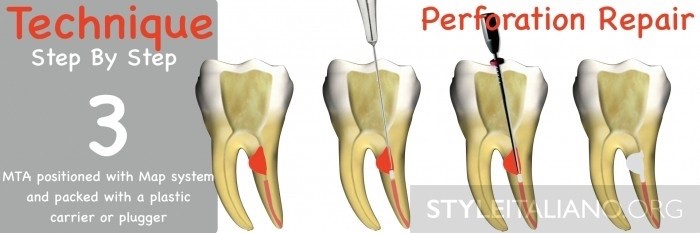
Fig. 4: During the third step MTA is positioned with MAP system. The system consists of a stainless steel or NiTi applicator with a bayonet catch for several exchangeable applicator cannulas (needles). Inside the cannula there is a plunger made in polymer that is longer than cannula providing a complete extrusion of internal material. The MTA can be taken from a dispenser thrusting the tip into the repair material and placed inside the canal in a sharp way pressing syringe piston to expel the material. An endo Micro Brush can be used to gently pack the MTA or a plastic carrier can be used for a stronger condensation. MTA must be placed to all the extension of perforation.
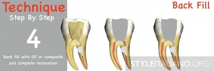
Fig. 5: The last step is represented by backfilling the coronal part with GP or composite material in a second visit.
Female patient, 47 years old, presented with a clinical picture of extensive iatrogenic perforation of the furcation region of the dental element 36 (Figs. ...
Combining Swiss precision and advanced engineering, the TruNatomy™ range offers clinicians a solution that provides efficient performance and increased ...
Align Technology announced the appointment of Angelo Maura as general manager for the MEA (Middle East and Africa) region. Bringing with him a wealth of ...
With Propex IQ®, Dentsply Sirona is now launching the first apex locator on the market that can be combined with a smart handpiece – the X-Smart IQ® –...
Today, dental treatment provided to animals is also performed using the latest technologies. Saving the teeth of animals is essential for basic needs such ...
To make the wait for ROOTS SUMMIT 2024 a little sweeter, the organisers would like to spotlight some of the speakers for next year’s edition, which will ...
INDIANAPOLIS, US: Though there have been many studies on the survival rate of endodontically treated teeth, only a relatively small proportion of these have...
A new method of detecting bacteria during root canal treatments could eradicate the need for follow up appointments and prevent treatments from failing, ...
The EssenSeal® is a new generation zinc oxide eugenol sealer featuring tea tree essential oil (Melaleuca) and composed of a powder base and a liquid ...
Dubai, United Arab Emirates: Clear aligners seem to be the best fit for patients’ lifestyles to align their teeth and improve their smiles. The Invisalign...
Education
Live webinar Tue. 17 March 2026 4:00 pm UAE (Dubai)
Held in January for the fourth time, Dentsply Sirona World Dubai has established itself as Dentsply Sirona’s flagship event for the Middle East, bringing ...
SHARJAH, UAE: Besides the negative side effects, health experts are increasingly concerned that long-term or improper use of chlorhexidine-containing ...
DAMMAM, Saudi Arabia: Aesthetic longevity remains a central concern in anterior composite restorations, where colour change is a frequent reason for ...

 International / International
 Brazil / Brasil
 Canada / Canada
 Latin America / Latinoamérica
 USA / USA
 Austria / Österreich
 Bosnia and Herzegovina / Босна и Херцеговина
 Bulgaria / България
 Croatia / Hrvatska
 Czech Republic & Slovakia / Česká republika & Slovensko
 France / France
 Germany / Deutschland
 Greece / ΕΛΛΑΔΑ
 Hungary / Hungary
 Italy / Italia
 Netherlands / Nederland
 Nordic / Nordic
 Poland / Polska
 Portugal / Portugal
 Romania & Moldova / România & Moldova
 Slovenia / Slovenija
 Serbia & Montenegro / Србија и Црна Гора
 Spain / España
 Switzerland / Schweiz
 Turkey / Türkiye
 UK & Ireland / UK & Ireland
 China / 中国
 India / भारत गणराज्य
 Pakistan / Pākistān
 Vietnam / Việt Nam
 ASEAN / ASEAN
 Israel / מְדִינַת יִשְׂרָאֵל
 Algeria, Morocco & Tunisia / الجزائر والمغرب وتونس

 Jovita Lawrence D'souzaRegister now1CELive webinar

 Dr. Giuseppe Luongo MD, DDS, Dr. Fabrizia Luongo DMD, MSRegister now1CELive webinar
 Dr. Stephanie Tran DDSRegister now1CELive webinar
 Dr. Alejandro LanisRegister now1CE


To post a reply please login or register