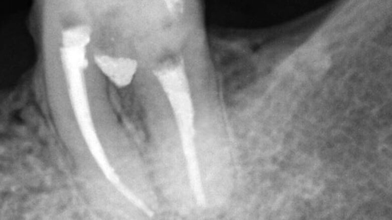
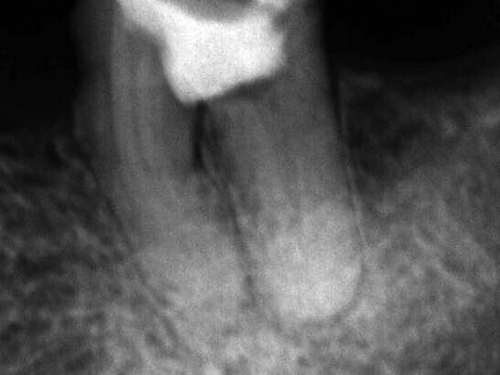
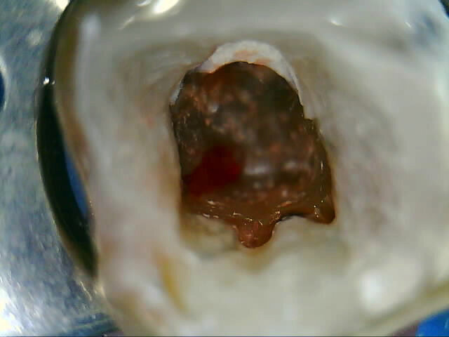
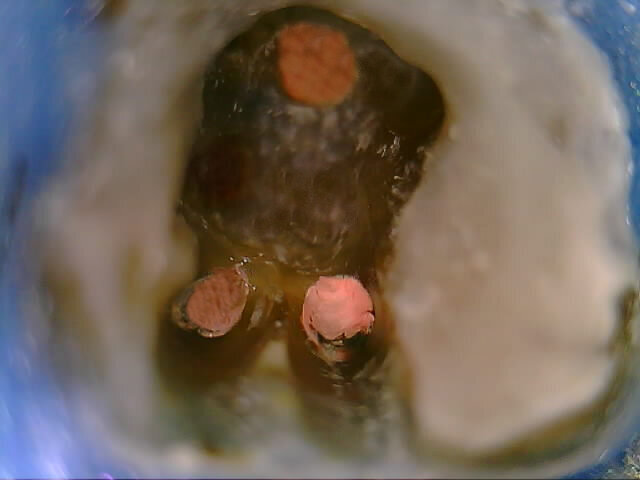
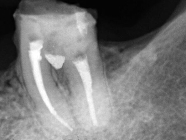
Female patient, 47 years old, presented with a clinical picture of extensive iatrogenic perforation of the furcation region of the dental element 36 (Figs. 1 and 2), associated with radiographic bone loss, vestibular fistula and pain on palpation. The patient reported history of having been previously subjected to an urgent intervention in this tooth by other professional, as it presented acute pain characteristic of pulpitis.
The tooth was submitted to endodontic therapy, and after the initial approach with the patient, anesthesia was given, followed by preparing absolute isolation. Subsequently, the coronary access was performed, where it was possible to clinically verify the presence of pulp necrosis and perforation. A disinfecting penetration of root canals (crown-down) was performed using as irrigator agent NaOCl to 5%, and the odontometry determined by the use of foraminal locator. The preparation was carried out by Reciproc system (VDW/Germany), and as irrigator agent was employed NaOCl 2.5% associated with ultrasonic activation performed with straight inserts (Irrisonic/Helse/Brazil).
Next, the drilling was treated, with its cleaning and the regularization, employing ultrasonic diamond insert (E7D/Helse/Brazil). As a complement to the intra-channel decontamination process and the furcation region, a biweekly exchange of Calcium Hydroxide (Ultracal/Ultradent/USA) was held, observing remission of all symptoms.
Fig. 1: Initial clinical and radiographic appearance of teeth 36
Fig. 2: Initial clinical and radiographic appearance of teeth 36
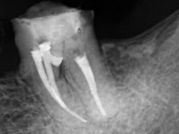
Fig. 3: Obturation of root canals.
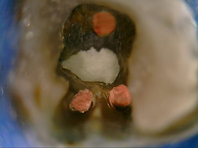
Figs. 4: Clinical and radiographic appearance of drilling filling with MTA Repair HP.
Figs. 5: Clinical and radiographic appearance of drilling filling with MTA Repair HP.
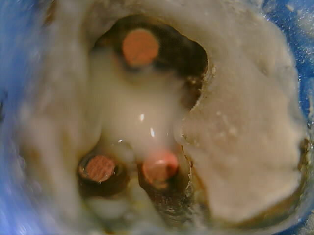
Fig. 6: Drilling region protection sealed with glass ionomer cement.
Fig. 7: Follow-up X-ray after two months.
The obturation was performed using the thermomechanical Hybrid Tagger technique (Fig. 3), by employing GutaCondensor (Maillefer/Switzerland), TP gutta-percha cones (Dentsply/Brazil) and MTA-based sealer Fillapex (Angelus/Brazil) (Fig. 4). After thermo compaction, the obturation cutting was performed, as well as vertical condensation using cold pusher; and again the region of the perforation was cleaned and filled with Calcium Hydroxide.
After 15 days, again, we proceeded to seal the drilled region, and initially verified the proper possibility of drying the area. The filling of the drilled region was carried out with the use of MTA Repair HP (Angelus/Brazil), previously prepared as recommended by the manufacturer, and it was inserted using an MTA Applicator (Angelus/Brazil). Clinical and radiographic criteria were used to determine the correct filling using the material (Figs. 4 and 5); and the glass ionomer cement (Vitremer/3M/USA) used for the protection of the sealed region (Fig. 6). After the temporary restoration, radiographically it was observed proper sealing of furcation region by MTA Repair HP, as well as no postoperative complications.
Follow up was conducted after two months, observing bone neoformation in the furcation region and absence of symptoms (Fig. 7).
Adjacent teeth damaged by dental bur
A clinical case that explains the technique step by step
In this article Dr. Thomas Zumstein presents a Neoss implant case that has been in function for more than 11 years. The case shows that long-term success ...
Molar incisor hypomineralisation (MIH) is a relatively common dental defect that appears in first permanent molars and incisors and varies in clinical ...
Every year, dental undergraduate and graduate students, with less than 2 years of clinical practice, are invited to participate by documenting a patient ...
A 50-year-old male patient presented for professional tooth cleaning. He had deep erosive lesions affecting the vestibular aspects of his maxillary central ...
The Smile Creator module is exocad’s advanced smile design solution for predictable aesthetic makeovers. Integrated into exocad’s DentalCAD platform, it...
As an oral surgeon and prosthodontist specialising in full-mouth rehabilitation, I have consistently sought to integrate advanced technologies into my ...
Implant dentistry has evolved dramatically in the 50 years since Branemark’s first patient was treated. The combination of improved micro-roughened ...
Worldwide, dental practitioners are convinced of this innovative, efficient latest generation bulk composite. Overwhelming feedback since its launch only ...
Live webinar
Tue. 24 February 2026
10:00 pm UAE (Dubai)
Prof. Dr. Markus B. Hürzeler
Live webinar
Wed. 25 February 2026
12:00 am UAE (Dubai)
Prof. Dr. Marcel A. Wainwright DDS, PhD
Live webinar
Wed. 25 February 2026
8:00 pm UAE (Dubai)
Prof. Dr. Daniel Edelhoff
Live webinar
Wed. 25 February 2026
10:00 pm UAE (Dubai)
Live webinar
Thu. 26 February 2026
5:00 am UAE (Dubai)
Live webinar
Tue. 3 March 2026
8:00 pm UAE (Dubai)
Dr. Omar Lugo Cirujano Maxilofacial
Live webinar
Wed. 4 March 2026
5:00 am UAE (Dubai)
Dr. Vasiliki Maseli DDS, MS, EdM


 Austria / Österreich
 Bosnia and Herzegovina / Босна и Херцеговина
 Bulgaria / България
 Croatia / Hrvatska
 Czech Republic & Slovakia / Česká republika & Slovensko
 France / France
 Germany / Deutschland
 Greece / ΕΛΛΑΔΑ
 Hungary / Hungary
 Italy / Italia
 Netherlands / Nederland
 Nordic / Nordic
 Poland / Polska
 Portugal / Portugal
 Romania & Moldova / România & Moldova
 Slovenia / Slovenija
 Serbia & Montenegro / Србија и Црна Гора
 Spain / España
 Switzerland / Schweiz
 Turkey / Türkiye
 UK & Ireland / UK & Ireland
 International / International
 Brazil / Brasil
 Canada / Canada
 Latin America / Latinoamérica
 USA / USA
 China / 中国
 India / भारत गणराज्य
 Pakistan / Pākistān
 Vietnam / Việt Nam
 ASEAN / ASEAN
 Israel / מְדִינַת יִשְׂרָאֵל
 Algeria, Morocco & Tunisia / الجزائر والمغرب وتونس










To post a reply please login or register